Summary
The safety and accuracy of laser eye surgery and lens-based vision correction have improved dramatically over the past decade, and the technology driving this improvement continues to advance in 2026. Patients choosing where to have surgery are increasingly aware that not all clinics use the same diagnostic tools or laser platforms. This article explains the key technologies used in modern ophthalmic assessment and surgery, what they measure, why they improve outcomes, and how the advanced diagnostic capability at Sussex Eye Laser Clinic directly benefits patient safety and visual results.
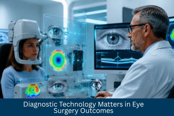
Why Diagnostic Technology Determines Surgical Outcomes
The precision of any vision correction procedure is ultimately limited by the quality of the measurements taken beforehand. A laser treatment plan is only as accurate as the pre-operative data fed into it. Corneal mapping, wavefront measurement, pupil tracking, and biometric calculation have all evolved significantly, and clinics that invest in the current generation of diagnostics are able to offer treatment that was not possible a decade ago.
Prof. Mayank Nanavaty at Sussex Eye Laser Clinic uses advanced diagnostic platforms to assess every patient before a treatment plan is finalised. Understanding what these tools measure helps patients appreciate why thorough pre-operative assessment is essential and why the assessment appointment is as important as the surgery itself.
Corneal Topography and Tomography
Corneal topography maps the surface curvature of the front of the cornea across thousands of points. It creates a detailed colour map of how the corneal surface curves in every meridian, identifying regular astigmatism, irregular astigmatism, and surface abnormalities.
Corneal tomography goes further. It measures both the front and back surfaces of the cornea and calculates the full three-dimensional corneal structure, including thickness at every point. This additional depth of information is critical for two reasons: it identifies very early keratoconus or forme fruste keratoconus that would be invisible on topography alone, and it allows precise calculation of how much corneal tissue will remain after laser treatment, ensuring the residual bed thickness is within the safety margin.
The Pentacam and similar tomography platforms used in modern pre-operative assessment provide more information about corneal safety than previous generation devices, and their use has substantially reduced the incidence of post-laser ectasia, which is one of the most serious potential complications of refractive surgery.
Wavefront Analysis
The optical system of the eye is not perfect. Beyond the common prescriptions of myopia, hyperopia, and astigmatism, which are called lower-order aberrations, the eye also has subtle optical imperfections called higher-order aberrations. These include spherical aberration, coma, and trefoil, among others. Higher-order aberrations contribute to symptoms such as halos around lights at night, glare, and reduced contrast sensitivity.
Wavefront analysis measures the full optical fingerprint of the eye, capturing both lower and higher-order aberrations in a single measurement. Wavefront-guided laser treatment uses this full optical map to customise the treatment profile specifically to the individual eye, rather than correcting only the prescription number. The result is a treatment that not only corrects the prescription but preserves or improves the optical quality of the eye compared to a standard treatment.
Wavefront-optimised treatment is a related but distinct approach. Rather than measuring and correcting existing higher-order aberrations, it uses a treatment profile designed to avoid introducing new aberrations from the surgery itself. Both approaches represent a significant improvement over older, non-customised laser treatments.
Eye Tracking During Laser Treatment
During a laser treatment that lasts only seconds, the eye makes tiny involuntary movements that, if not accounted for, could displace the ablation from its intended centre. Modern laser platforms use high-speed eye-tracking at 1,000 or more measurements per second, continuously monitoring the position of the pupil and adjusting the laser delivery to follow it in real time.
Advanced systems also track torsional rotation, the subtle rotation of the eye around its visual axis that occurs when a patient moves from an upright sitting position to lying flat under the laser. Compensating for this rotation is particularly important for the accurate correction of astigmatism, where the axis of the cylinder must be applied precisely.
Optical Biometry for Intraocular Lens Calculation
For patients undergoing cataract surgery or refractive lens exchange, the most important calculation is the power of the intraocular lens that will be implanted. Modern optical biometry measures the axial length of the eye, the curvature of the anterior cornea, the depth of the anterior chamber, and the lens thickness using optical coherence interferometry, which is highly accurate and completely non-contact.
These measurements feed into advanced intraocular lens power formulae, the most modern of which account for the individual anatomy of the eye in ways that significantly reduce the residual prescription after surgery. The result is more patients achieving spectacle independence after lens surgery than was possible with older measurement and calculation methods.
Testimonials
“Excellent manner, listens really well and gives multiple opportunities to ask questions, which is really helpful. Very calm, gently spoken and not rushed”
The Role of Artificial Intelligence in Ophthalmic Diagnostics
Artificial intelligence is increasingly integrated into ophthalmic diagnostic platforms. AI-based analysis of corneal tomography data can identify subtle patterns associated with early keratoconus with higher sensitivity than human review of the same data. AI-assisted IOL power calculation incorporates large datasets of post-operative outcomes to improve prediction accuracy for eyes that fall outside the parameters of traditional formulae.
In screening, AI tools are being validated for the detection of diabetic retinopathy and macular degeneration from retinal photographs, a development that has the potential to extend the reach of specialist screening to a far wider population. While AI does not replace the clinical judgement of a consultant ophthalmologist, it is an increasingly powerful tool for improving the consistency and accuracy of the diagnostic process.
Conclusion
Technology does not perform surgery. An experienced, fellowship-trained ophthalmic surgeon with excellent clinical judgement remains the essential element of a safe and successful outcome. What modern diagnostic technology does is provide that surgeon with better, more complete information on which to base every decision. At Sussex Eye Laser Clinic, Prof. Mayank Nanavaty combines consultant-level clinical expertise with access to advanced diagnostic platforms to provide the quality of assessment that underpins consistently excellent surgical outcomes. Book a consultation at sussexeyelaserclinic.co.uk.
Frequently Asked Questions
What is the difference between wavefront-guided and wavefront-optimised laser treatment?
Wavefront-guided treatment measures the existing higher-order aberrations of the individual eye and designs a treatment profile specifically to correct them. Wavefront-optimised treatment uses a standardised profile designed to avoid introducing new higher-order aberrations from the surgery, without attempting to correct existing ones. Both are superior to standard laser treatment. The appropriate choice depends on the individual’s aberration profile and is discussed at consultation.
How long does a pre-operative assessment appointment take at Sussex Eye Laser Clinic?
A comprehensive pre-operative assessment typically takes between 90 minutes and two hours. This allows time for corneal tomography, wavefront analysis, biometry, pupil measurement, tear film assessment, and a thorough consultation with Prof. Nanavaty to discuss the findings, the recommended treatment, and to answer all questions.
Why does corneal tomography reduce the risk of complications?
Corneal tomography maps the full three-dimensional structure of the cornea, including areas of thinning or irregular curvature that standard topography does not capture. This allows the identification of eyes that are at elevated risk of post-laser ectasia, a thinning and bulging of the cornea that can occur if an inappropriate eye is treated. By identifying these eyes before surgery, corneal tomography is the primary reason that serious post-laser complications are now rare at well-equipped clinics.