Keratoconus is a progressive eye condition that can quietly steal a person’s visual clarity during their most formative years. For decades, the medical community viewed the bulging, cone-like deformation of the cornea as an inevitable slide toward major disability or invasive surgery. However, the advent of Corneal Collagen Crosslinking has fundamentally shifted the treatment paradigm. Instead of waiting for the cornea to fail, doctors can now intervene early to reinforce the eye’s structural integrity. By understanding how this procedure works and recognizing the early warning signs, patients can protect their sight and maintain a high quality of life.
Corneal Collagen Strengthening: Reinforcing the "Glue" of the Eye
To understand why Corneal Collagen Crosslinking is so effective, one must first understand the anatomy of the cornea. The cornea is the clear, dome-shaped front surface of your eye, composed largely of layers of corneal collagen fibers. In a healthy eye, these fibers are anchored together by chemical “cross-links” that act like invisible biological glue, keeping the cornea stiff enough to maintain its round shape against the internal pressure of the eye.
In a patient with keratoconus, these collagen bonds are abnormally weak or sparse. As a result, the cornea loses its structural rigidity and begins to bulge outward into a cone shape. This irregularity causes light to scatter as it enters the eye, leading to blurred vision and high levels of astigmatism.
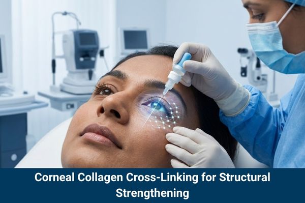
Corneal Collagen Cross-linking solves this problem through a sophisticated biochemical reaction. During the procedure, a Corneal Cross-linking Specialist applies a specialized solution of Riboflavin (Vitamin B2) to the eye. Once the cornea is saturated, it is exposed to a precisely controlled amount of Ultraviolet-A (UVA) light. This interaction triggers the release of reactive oxygen species that create new, strong covalent bonds between the existing corneal collagen strands. By essentially “re-gluing” the eye at a molecular level, the cornea becomes significantly stiffer and more resistant to further bulging.
Read Our Article
When Children Inherit Weak Eyes: Can Early CXL Protect Their Future?Corneal Crosslinking Specialist Insights: Red Flags for Early Action
In the world of progressive eye disease, timing is the most critical factor. Because corneal cross linking is designed to stop progression rather than reverse existing damage, identifying the disease in its earliest stages is the key to a successful outcome. This is why a consultation with a Corneal Crosslinking Specialist is vital for anyone experiencing rapid changes in their vision.
Modern diagnostic technology has revolutionized our ability to spot these early “red flags.” Specialists now use advanced corneal topography and tomography – high-tech mapping systems that create a 3D “GPS map” of the eye’s surface and thickness. These scans can detect microscopic thinning and subtle steepening of the corneal curve long before a patient notices a significant drop in their vision.
Key indicators that signal the need for immediate Corneal Crosslinking include:
- Frequent Prescription Changes:Needing a new pair of glasses or contacts every few months due to increasing astigmatism.
- Corneal Thinning:Measurements showing the corneal wall is losing its thickness.
- Ghosting or Halos:Seeing “double” or “smearing” around lights, even with corrective lenses.
By intervening the moment these progressive changes are documented, patients can “lock” their vision in place, preventing the permanent scarring that often occurs in later stages.
Corneal Crosslinking Outcomes: Halting the "Blur" for Visual Stability
One of the most important conversations a Corneal Cross-linking Specialist has with their patient involves setting realistic expectations. It is vital to understand that the primary goal of this corneal surgery is stabilization, not necessarily a total cure or the achievement of 20/20 vision without glasses.
The “win” in CXL therapy is a stable map of the eye. For the vast majority of patients, Corneal Crosslinking successfully halts the progression of keratoconus, meaning their vision stops getting worse. In a world where keratoconus naturally declines over time, “staying the same” is a clinical victory.
However, there is an added benefit for many. Clinical data suggests that approximately 30–40% of patients experience a slight “flattening” effect of the corneal cone in the years following the procedure. While this might not eliminate the need for glasses, it often makes the eye’s shape more regular. This “regularization” can make it much easier and more comfortable to fit contact lenses – especially specialized scleral lenses – and can sometimes result in a modest improvement in best-corrected visual acuity.
Testimonials
“Extremely gentle, professional and informative, Could not wish for more”
Corneal Collagen Cross-linking for Teens: Specialized Care for Young Patients
Keratoconus is notoriously aggressive in children and teenagers. Because the young eye is still developing and the corneal collagen is naturally more flexible, the rate of deterioration in a 15-year-old can be significantly faster than in a 40-year-old. For this reason, many experts have moved away from the traditional “wait and see” approach for pediatric patients.
When a teenager is diagnosed, a Corneal Cross-linking Specialist will often recommend moving straight to treatment. Waiting six months to “confirm” progression in a teen could result in a permanent loss of functional vision that might have been saved with earlier intervention.
By prioritizing Corneal Collagen Cross-linking in young patients, we ensure that they can continue their education, participate in sports and eventually drive without the looming threat of legal blindness. Early treatment protects their future career options and reduces the psychological burden that comes with a chronic, deteriorating condition during their most social years.
Our Other Treatments
Laser Refractive Eye SurgeryCorneal Transplant Surgery Prevention: Avoiding Major Invasive Procedures
Before the FDA approval and widespread adoption of CXL, the endgame for many keratoconus patients was a corneal transplant surgery. While transplants are a marvel of modern medicine, they are considered a last resort. Any corneal surgery involving a donor tissue transplant carries risks, including graft rejection, infection and a lifetime requirement for steroid eye drops.
Corneal Collagen Crosslinking has fundamentally changed this trajectory. Longitudinal studies now show that the success rates for CXL are incredibly high, with stabilization occurring in over 90% of cases. By strengthening the cornea early on, we can effectively “cancel” the need for high-risk corneal transplant surgery later in life.
Avoiding a transplant is not just about avoiding the operating room; it’s about maintaining the patient’s own biological tissue. A natural cornea, reinforced by cross-linking, is almost always superior in terms of long-term health and visual predictability than a donor graft. For the patient, this means fewer follow-up surgeries, lower costs over their lifetime and a much lower risk of sudden vision-threatening complications.
Corneal Crosslinking Recovery: What to Expect During Life After CXL
The recovery process following Corneal Collagen Crosslinking is relatively brief but requires diligence. Because the procedure involves removing the thin outer layer of the cornea (in the “Epi-Off” technique) to allow the Riboflavin to soak in, the first few days are focused on epithelial healing.
Immediately following the corneal surgery, the doctor will place a clear “bandage” contact lens over the eye. This lens acts as a synthetic scab, protecting the surface while the new cells grow back. Patients typically use a regimen of antibiotic and anti-inflammatory steroid drops for several weeks to prevent infection and manage swelling.
It is common to experience some “corneal haze” in the first month or two. This is a slight cloudiness that occurs as the corneal collagen fibers settle into their new, stiffer arrangement. For most, this haze is temporary and clears up as the eye heals, eventually leaving the patient with a stable, stronger ocular surface. While the eye may feel scratchy or sensitive to light for the first 48 to 72 hours, most patients return to their normal routines – and their specialized contact lens fittings – within a few weeks.
Conclusion
The journey with keratoconus no longer has to be one of fear and uncertainty. Thanks to Corneal Collagen Crosslinking, we have moved from a reactive “wait for failure” model to a proactive “preventative” model. By reinforcing the corneal collagen at the first sign of trouble, patients can halt the progression of their disease and avoid the complexities of corneal transplant surgery.
If you or a loved one are noticing the “red flags” of distorted vision or frequent prescription changes, the best course of action is to consult with a Corneal Crosslinking Specialist immediately. Early intervention is the most powerful tool we have to preserve sight, ensuring that the world remains clear and focused for years to come.